
Dental imaging is how we find underlying oral problems before they become serious — and how we plan complex clinical treatments with absolute precision. At Crystal River Dental, we use modern digital technology that delivers diagnostic clarity with a fraction of the radiation of older systems.
Standard visual screenings only reveal surface enamel health. Critical underlying dynamic concerns — including deep decay patterns hiding between tight teeth, early jawbone reabsorption tracks, hidden root abscesses, impacted third molars, cysts, and tumor paths — stay completely concealed beneath structural surfaces. Dental radiography isn’t an elective operational add-on; it is an irreplaceable baseline patient protection mechanism. We frame diagnostics strictly around preventative security, never revenue generation fields.
Full Scope Checklist
Tactile examination checking for decay, fracture lines, enamel attrition wear, and aging restoration integrity status fields.
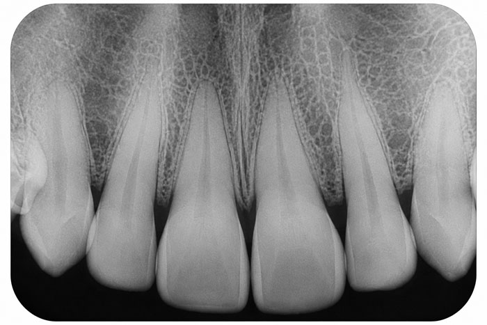
Displays an entire singular tooth profile from top crown down to the root tip anchor bone density. Used to verify root canal infections or absolute fracture paths.
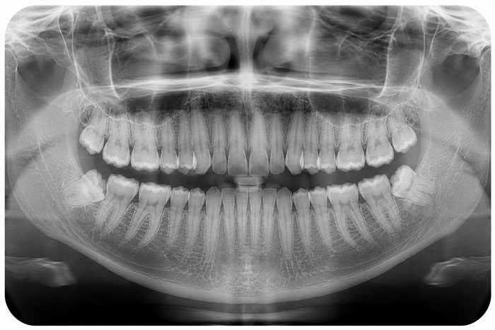
Generates a single wide-view flat map layout of the entire maxilla and mandible arches, jaw joints (TMJ), and sinus floors. Vital for wisdom teeth mapping.
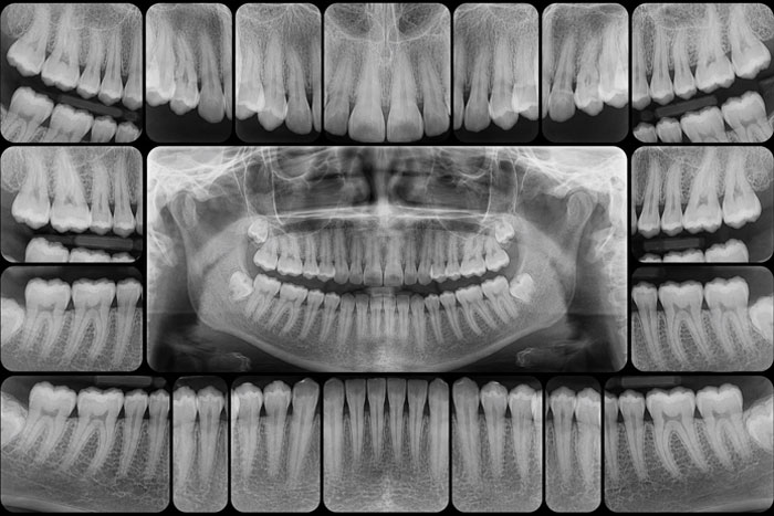
A comprehensive structural set of 14–20 individual localized digital sensors. Captures the complete baseline diagnostic grid for incoming guest charts.
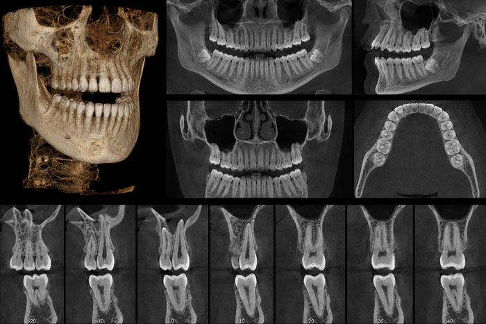
Computes a highly accurate 3D volumetric model of bone thickness, nerve networks, and vascular pathways. Critical for zero-compromise surgical implant plotting.
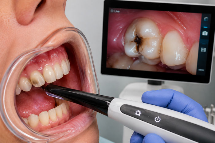
A pen-sized micro-wand lens displaying high-definition live color video of internal tooth surface fractures or leakage margins right onto your chairside monitor.
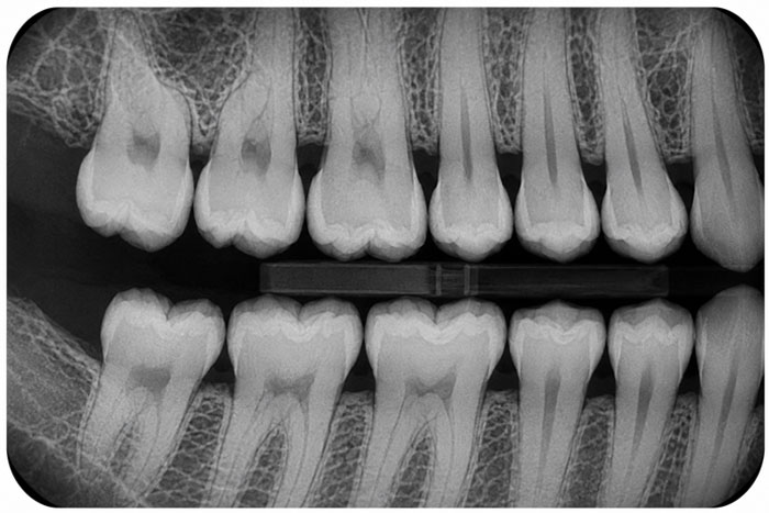
For healthy adult recall patients, localized digital bitewing checks are suggested every 12 to 24 months. New patients typically require a full mouth series or structural panoramic scan to anchor an accurate, baseline tracking metric.
Yes. Because digital capture arrays cut down output thresholds by up to 90% compared to legacy film layers, radiation exposure is remarkably nominal. When combined with our protective lead apron layering, the procedure is verified safe across children and expectant mothers.
While we always honor your ultimate treatment autonomy, declining necessary diagnostic imaging completely compromises our clinical capacity to screen for sub-surface path trends or safely clear teeth for clinical adjustments. We document your parameters meticulously and work within safety parameters.
Cone Beam Computed Tomography rotates a specialized sensor array to capture a precise 3D rendering of bone depth, neural paths, and sinus layouts. It is universally required for accurate surgical implant design, complex wisdom extractions, and advanced airway evaluations.
Standard basic diagnostic images like bitewings or baseline panoramic scans are almost universally covered fully (at 80% to 100%) within preventive insurance allowances. High-tier surgical 3D CBCT scans can occasionally necessitate pre-authorization.
High-resolution 3D CBCT mapping generates accurate structural data fields, securing safe, predictable prosthetic implant placements.